Thermo Fisher Scientific Logo
      Video home
      close menu
      Video home
      close
      • Home
      • Antibodies Videos
      • Bioprocessing Videos
      • Cancer Research Videos
      • Cell Culture Videos
      • Cell & Gene Therapy Videos
      • Clinical Microbiology Videos
      • Digital Science Videos
      • DNA & RNA Extraction & Analysis Videos
      • Electron Microscopy
      • Environmental Videos
      • Food and Beverage Videos
      • Genome Editing Videos
      • Manufacturing & Processing Videos
      • Mass Spectrometry Videos
      • PCR Videos
      • Protein Biology Videos
      • Radiation Detection & Measurement Videos
      • Real-Time PCR Videos
      • Sequencing Videos
      • Spectroscopy, Elemental & Isotope Analysis Videos
      • Stem Cell Research

      Step-by-step guide to successful western blot analysis

      3:05

      • Share on Facebook
      • Tweet this video
      • Share on LinkedIn
      • Share via Email
      Description
      Related Videos

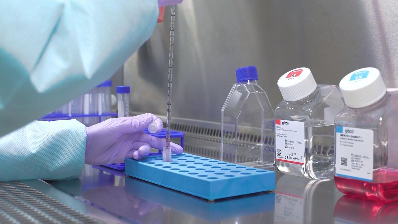
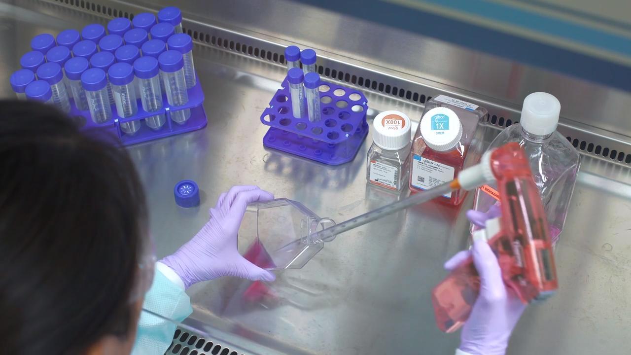
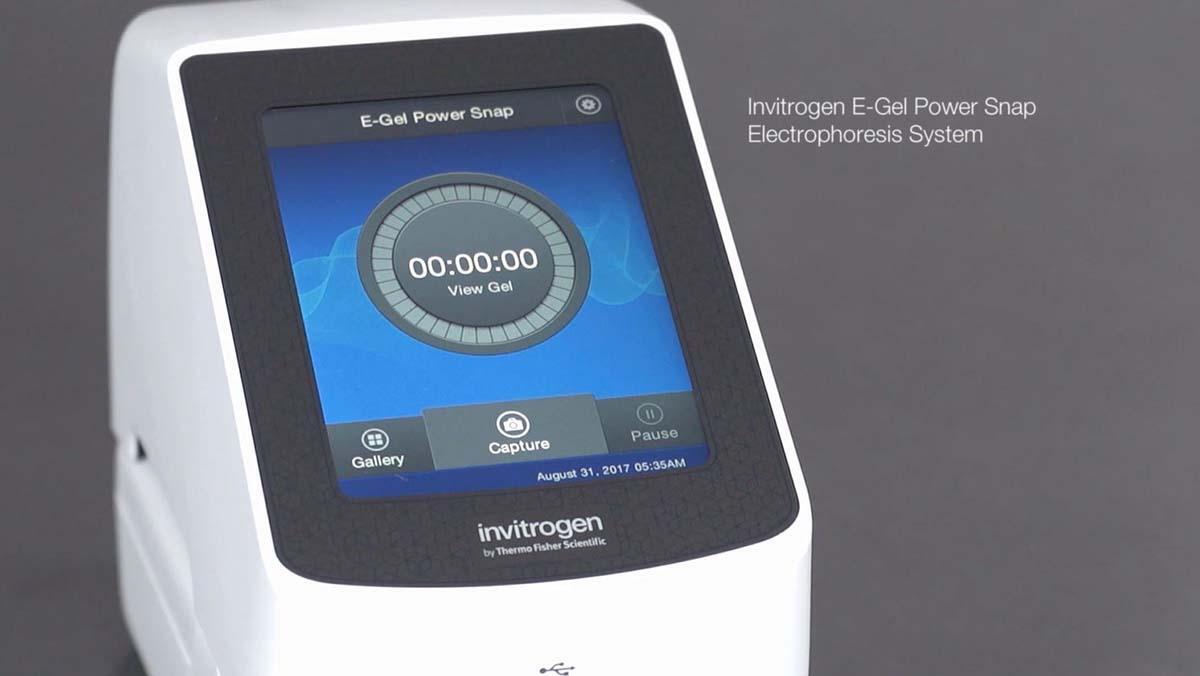
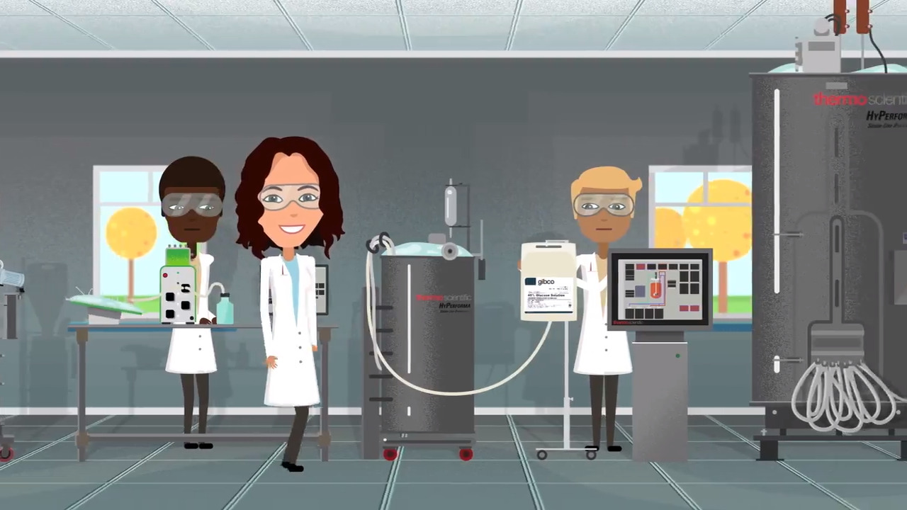
      Western Blotting is the most widely used technique for protein detection. In this video, we will cover the 3 major steps in generating a western blot: Separate, Transfer, and Detect. After protein samples have been prepared, the mixture is separated by molecular weight using protein gel electrophoresis, which is a standard laboratory technique by which charged protein molecules are transported through a matrix by an electrical field. Once the proteins have been separated in the gel, they must be transferred to a solid support membrane, which facilitates protein detection through the use of antibodies specific to the target protein. Using a traditional apparatus, the protein transfer process can take 30 to 60 minutes to complete. However, new technologies have been developed that can cut the transfer time down to as little as 7 minutes. Once complete, the efficiency of the transfer can be confirmed by staining the membrane with a reversible stain. The membrane is now ready for detection. This is a multi-step process, and the first step is minimizing nonspecific binding by using a blocking agent. To do this the membrane should first be incubated with a protein containing solution such as nonfat dry milk or a purified protein like bovine serum albumin, or BSA. Proteins in this blocking solution occupy the empty spaces on the membrane to help prevent detection antibodies in the next step from sticking to the membrane. Recent advancements in blocking agents have helped make this step faster and more efficient. In this step, the membrane is incubated with the primary antibody specific to the target to allow the antibody to bind its target. Any unbound primary antibody is washed away. A secondary antibody that is specific to the primary antibody is then added. The secondary antibody is typically tagged with a reporter enzyme like Horse Radish Peroxidase or a fluorophore. As before, any unbound secondary antibody is washed away. If using an enzyme-based system, a few different substrates can be used, which produce either a colorimetric or chemiluminescence signal. Chemiluminescence detection is the method of choice in many protein laboratories, as it provides high sensitivity and convenience for detection with a film or digital imaging equipment. Today more researchers are taking advantage of fluorescently conjugated secondary antibodies that eliminate the need for substrates and provides the ability to detect multiple proteins at the same time. Western blot imaging and documentation can be performed using an X-ray film for chemiluminescence detection, or increasingly with western blot imaging systems equipped with charge-coupled device cameras. In summary, the western workflow consists of 3 major steps: 1. Separate proteins by gel electrophoresis 2. Transfer proteins from the gel to a solid support and 3. Detect, where we use antibodies specific to the target protein to visualize the protein of interest Depending on your experimental needs, there are reagents and equipment that can help optimize every step of the western blot process. Find out more about western blotting at thermofisher.com/western.

      View More
      View Less
      Learn more

      Share

      • Share on Facebook
      • Tweet this video
      • Share on LinkedIn
      • Share via Email

      Related Videos

      In Popular videos

      • Play video How to separate proteins using an Invitrogen precast SDS-PAGE gel

        How to separate proteins using an Invitrogen precast SDS-PAGE gel

        Learn how to separate proteins by using a Invitrogen Bolt Bis-Tris Plus SDS-PAGE precast gel and Mini Gel Tank

        5:24

      • Play video Freezing Cells: Cell Culture Basics

        Freezing Cells: Cell Culture Basics

        This cell culture workflow video focuses on steps pertaining to freezing cells. It also contains useful information, troubleshooting guides and protocols for the experienced researcher as well.

        4:52

      • Play video Thawing Cells: Cell Culture Basics

        Thawing Cells: Cell Culture Basics

        This video provides an introduction to cell culture, with a focus on maintaining cell health throughout the processes of culturing, freezing, thawing and passaging cells. In this video, we focus on how to thaw cells.

        3:57

      • Play video Quantify DNA, RNA, or protein with the Qubit 4 Fluorometer

        Quantify DNA, RNA, or protein with the Qubit 4 Fluorometer

        Quickly quantitate the amount of DNA, RNA, or protein for proper preparation of the next step in your experiment. The quantitation process is simple, fast and even includes a built-in Reagent Calculator.

        1:45

      • Play video Invitrogen E-Gel Power Snap System Features

        Invitrogen E-Gel Power Snap System Features

        Learn about the E-Gel Power Snap System powerful features and how the integrated design combines the convenience of rapid, real-time nucleic acid analysis with high-resolution image capture to reduce workflow time and accelerate discovery.

        1:51

      • Play video What Makes a “Good” Bioprocess?

        What Makes a “Good” Bioprocess?

        The article introduces three main questions a manufacturer should ask themselves: 1) How do I simplify my process? 2) How do I ensure consistent performance? 3) How is upstream impacting downstream?

        1:04

      •  
      •  
      •  
      •  
      •  
      Brands
      • Thermo Scientific
      • Applied Biosystems
      • Invitrogen
      • Fisher Scientific
      • Unity Lab Services
      Shopping Tool
      • Product Selection Guides - US
      • Quick Order - US
      • Redeem a Quote
        * Please enter a valid quote
      • New Products
      • Promotions
      • Mobile & Desktop Apps
      • Shared Lists
      eSolutions
      • eProcurement
      • Supply Center
      • Instrument Management
      Support
      • Order Support
      • Training
      • Webinars
      • Blog
      • Social Media
      • Contact Us
      • Report a Site Issue
      Thermo Fisher Scientific
      • About Us
      • Careers
      • Investors
      • News
      • Responsibility
      • Trademarks
      United States
      • Terms & Conditions
      • Privacy Information Center
      • Price & Freight Policy


      Note: You clicked on an external link, which has been disabled in order to keep your shopping session open.

      Ok

      Share this video

      Embed

      Size: x pixels