Thermo Fisher Scientific Logo
      Video home
      close menu
      Video home
      close
      • Home
      • Antibodies Videos
      • Bioprocessing Videos
      • Cancer Research Videos
      • Cell Culture Videos
      • Cell & Gene Therapy Videos
      • Clinical Microbiology Videos
      • Digital Science Videos
      • DNA & RNA Extraction & Analysis Videos
      • Electron Microscopy
      • Environmental Videos
      • Food and Beverage Videos
      • Genome Editing Videos
      • Manufacturing & Processing Videos
      • Mass Spectrometry Videos
      • PCR Videos
      • Protein Biology Videos
      • Radiation Detection & Measurement Videos
      • Real-Time PCR Videos
      • Sequencing Videos
      • Spectroscopy, Elemental & Isotope Analysis Videos
      • Stem Cell Research

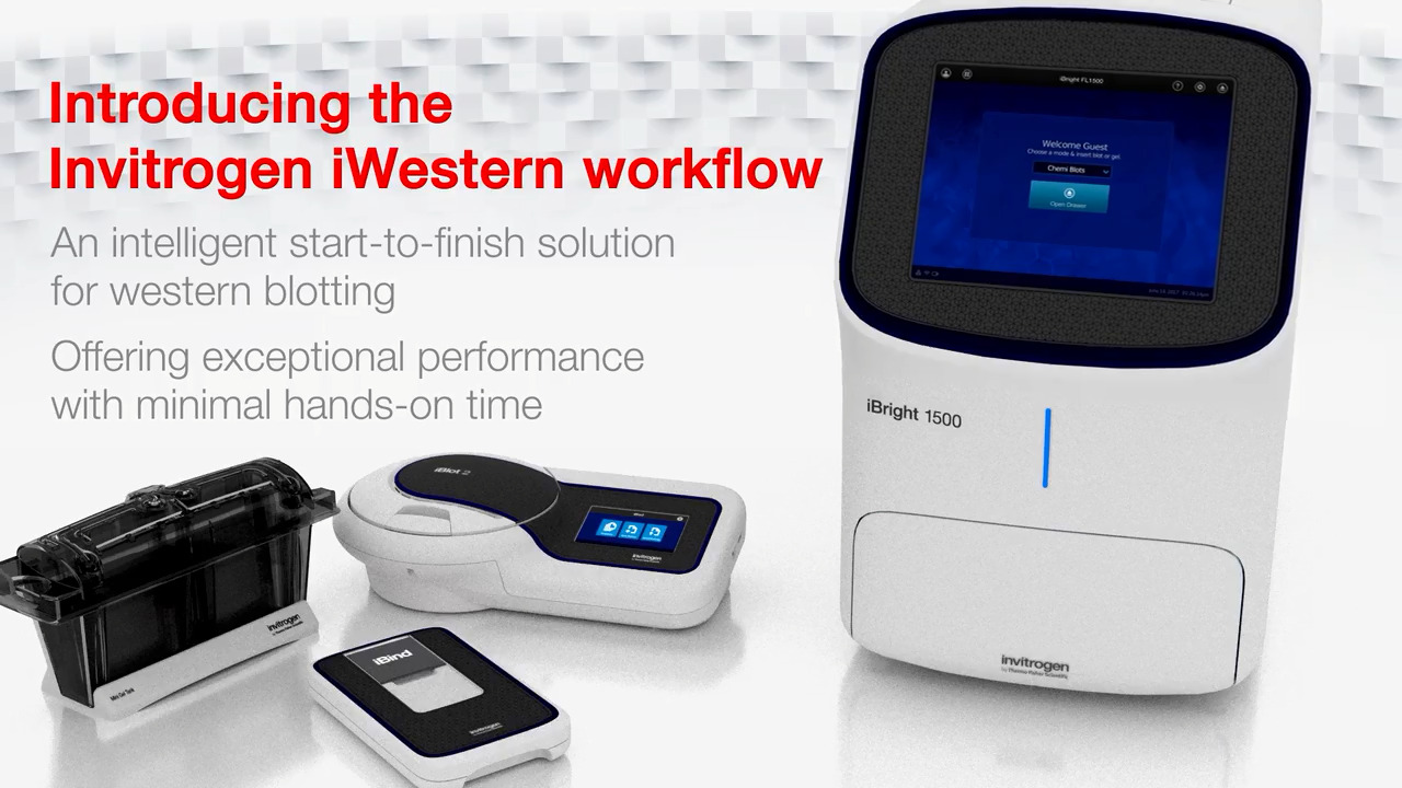
      How to perform a western wet tank transfer using the Invitrogen Mini Blot Module

      4:05

      • Share on Facebook
      • Share on X
      • Share on LinkedIn
      • Share via Email
      Description
      Related Videos

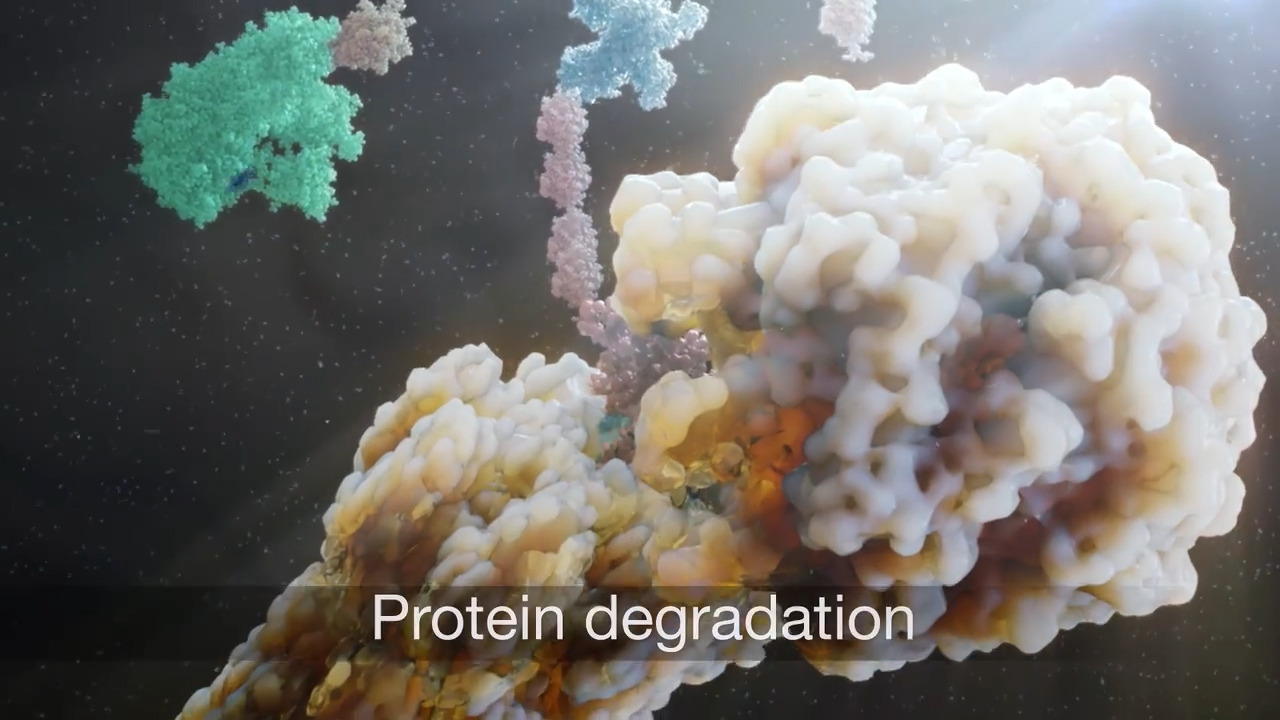
      Once the proteins have been separated in the gel, they now can be transferred to a solid support membrane to start the western blotting workflow. In this video, you’ll learn how to perform a western transfer using the Mini Blot module in a Mini Gel Tank. Make sure the cassette clamps are removed before you set up your transfers. One gel is transferred per module, and two modules fit in one tank. Included with the Mini Blot Module kit is a roller, sponge pads, tweezers, and a quick reference card. Approximately 250 milliliters of Transfer Buffer is required for each transfer. Prepare Transfer Buffer by adding 25 milliliters of 20X Bolt Transfer Buffer, 50 milliliters of methanol, and 500 microliter of Bolt Antioxidant, and bring the total volume up to 500 milliliters with deionized water. Soak two sponge pads in transfer Buffer and Squeeze the pads to remove air bubbles. Open the gel cassette using a gel knife and carefully remove the wells and the foot of the gel, so that the entire gel is of equal thickness. Place a shallow dish on the bench and pour 1X transfer buffer to dip the filter papers into during sandwich assembly. Assemble the western transfer sandwich by placing the cathode or negative core on a flat surface. Pour 5 milliliters of transfer buffer into the core, then place a pre-soaked sponge pad on the cathode core followed by a pre-wetted filter paper. Next, use the blot roller to squeeze out the air bubbles from between each layer. Place the gel on the stack, again using the roller to remove air bubbles. Using tweezers, place the membrane on top of the gel and roll once more. Complete the stack by layering another pre-wetted filter paper on top of the membrane, removing bubbles, and placing pre-wetted sponge pads on top of the filter paper. Complete the assembly by placing the anode core on top of the sandwich and pressing the two module halves together. Insert the blot module into the Mini Gel Tank with the cathode core facing the front. The blot module should be seated with the electrodes contacting the electrode bar. Extra transfer buffer can be added to the module core to submerge the sandwich, but don’t fill it above the gasket. Add transfer Buffer or deionized water—about 225 milliliters to the chamber, just below the electrode bar. Make sure the power supply is off. Place the cover on the tank and plug the power leads into the power supply. Turn the power supply on to begin the transfer. For Nitrocellulose membranes, transfer at a constant 10 volts for 60 minutes. For PVDF membranes, transfer at 20 volts for 60 minutes. After the transfer is complete, turn off the power supply, disconnect the cords, open the lid by placing your thumb on the tank handles pulling the lid up with your fingers. Remove the module, pour out the buffer, and disassemble the module. Remove the membrane using tweezers. You are now ready to probe your membrane for western analysis. Find out more information about western blotting with the Mini Blot Module at thermofisher.com/westerntransfer

      View More
      View Less
      Learn more

      Share

      • Share on Facebook
      • Share on X
      • Share on LinkedIn
      • Share via Email

      Related Videos

      In Protein Biology Videos

      • Play video Expanding ERF (Equivalent Number of Reference Fluorophore) Value Assignments for Quantitative Flow Cytometry- More Colors and Smaller Particles

        Expanding ERF (Equivalent Number of Reference Fluorophore) Value Assignments for Quantitative Flow Cytometry- More Colors and Smaller Particles

        We will introduce ERF (Equivalent Number of Reference Fluorophore) standards and discuss the significance and advantages of using the ERF standards for quantitative flow cytometry instead of pre-existing, value-assigned beads.

        52:22

      • Play video Advancing multiplex imaging for spatial proteomics research

        Advancing multiplex imaging for spatial proteomics research

        Learn about the Spatial Biology workflow at a high level and then understand how to image mIF with the EVOS S1000 Spatial Imaging System

        25:31

      • Play video Ask a protein pro - Tips to help you improve your western blotting: Antibody selection

        Ask a protein pro - Tips to help you improve your western blotting: Antibody selection

        In this video we discuss key considerations for selecting effective antibodies for your western blotting.

        5:21

      • Play video Invitrogen iBright Analysis Software – An introduction and overview of key analysis workflows

        Invitrogen iBright Analysis Software – An introduction and overview of key analysis workflows

        Learn how to navigate through the iBright Analysis Software interface, how to perform routine adjustments for gel and western blot images, and understand the basic analysis workflows.

        58:48

      • Play video Introduction to protein degradation

        Introduction to protein degradation

        This video will provide an overview of protein degradation, ubiquitin proteasome pathway (UPP), and targeted protein degradation. Learn more at http://thermofisher.com/proteindegradation

        3:08

      • Play video Quantitative western blotting analysis: tips & streamlining analysis using Invitrogen iBright Analysis Software

        Quantitative western blotting analysis: tips & streamlining analysis using Invitrogen iBright Analysis Software

        Gain insights to best practices and considerations that can improve the quality of western blots and subsequent quantitative data analysis.

        59:12

      •  
      •  
      •  
      •  
      •  
      Brands
      • Thermo Scientific
      • Applied Biosystems
      • Invitrogen
      • Fisher Scientific
      • Unity Lab Services
      Shopping Tool
      • Product Selection Guides - US
      • Quick Order - US
      • Redeem a Quote
        * Please enter a valid quote
      • New Products
      • Promotions
      • Mobile & Desktop Apps
      • Shared Lists
      eSolutions
      • eProcurement
      • Supply Center
      • Instrument Management
      Support
      • Order Support
      • Training
      • Webinars
      • Blog
      • Social Media
      • Contact Us
      • Report a Site Issue
      Thermo Fisher Scientific
      • About Us
      • Careers
      • Investors
      • News
      • Responsibility
      • Trademarks
      United States
      • Terms & Conditions
      • Privacy Information Center
      • Price & Freight Policy


      Note: You clicked on an external link, which has been disabled in order to keep your shopping session open.

      Ok

      Share this video

      Embed

      Size: x pixels